
Causes of Facial Asymmetry
Similar-looking facial differences can have different causes. Development, teeth, soft tissues, injury and facial movement all contribute to appearance. Start with when the difference began and whether function changed; a photograph alone cannot identify the cause.
When did you first notice the difference?
“My right cheek looks bigger” describes an appearance, not an explanation. First separate a newly noticed feature from a feature that has actually changed. An unfamiliar camera view can reveal a difference that was already present; a genuinely new difference deserves attention even if an older photo is unavailable.
Look for a history, not a verdict
Old photographs can help establish timing. Similarity to relatives may be useful background, but neither family resemblance nor long duration proves that a difference is harmless.
Describe the change
Note roughly when it began, where it is, and whether it persists away from the camera. A new or worsening difference should be assessed by a medical or dental professional.
Mention function
Tell a clinician about changes in smiling, eye closure, speech, chewing or sensation. A neutral portrait alone leaves out movement.
These observations organize a conversation. They cannot sort you into a diagnosis or rule out a condition.
One visible outline, several possible sources
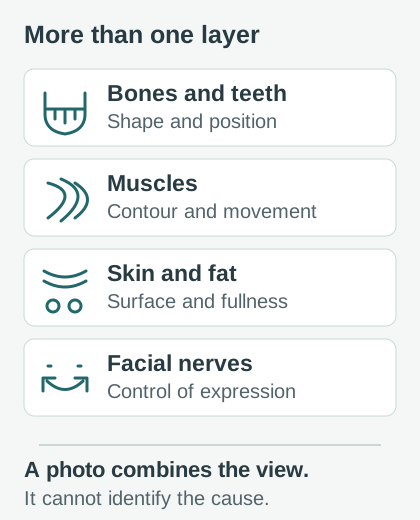
A face is not a single layer. Its outline combines the facial skeleton and teeth, muscle, fat, skin and other soft tissues. Facial nerves help control expression. A difference can involve more than one of these, and the same visible feature can have different explanations.
Penn Medicine’s overview describes developmental, injury-related, dental, muscular and neurological contributors. Identifying one requires history and examination; a cheek contour cannot reveal which tissue is responsible.
- Structural
- Differences in shape, size or position, such as an uneven jaw or tooth alignment.
- Functional
- Differences in how the face moves or works, such as a change in eye closure or chewing.
- Photographic
- Differences in the projected image, influenced by viewpoint, light and expression.
These descriptions can coexist. “Structural” does not mean permanent, and “functional” does not mean something to exercise without assessment.
Development, aging and injury do different things
Development does not make identical halves
Small differences are common. Some people also have a defined congenital condition. For example, hemifacial microsomia affects development of structures on one side of the face, often including the jaw and ear. This is one specific condition, not an explanation to assign to every uneven jaw.
An old photograph helps with the timeline. It cannot tell you whether a difference comes from bone, muscle or soft tissue.
Adult faces continue to change
A 2018 study of 191 volunteers found greater measured 3D asymmetry at older ages. This was a comparison across people, not a forecast for an individual. The findings do not justify assuming that a new change is “just aging.” Adult bone and soft tissues are not fixed.
Injury can alter bones, muscles or soft tissue. Mention prior facial injuries and procedures during an assessment, including the timing of any change. Their presence in your history does not by itself establish the cause.
Use symptoms to describe the problem clearly
Teeth and bite
Tooth position, missing teeth and how the jaws meet can affect the visible lower face. Report a change in your bite or difficulty chewing to a dentist. Do not assume that orthodontic treatment is required from a selfie.
Jaw joints and chewing muscles
TMJ names the joint; TMD names disorders affecting jaw joints and chewing muscles. Pain, locking or limited opening are useful symptoms to report. NIDCR explains that research does not support a bad bite or orthodontic treatment as a cause of TMD. Tooth alignment and TMD are separate questions.
Nerves and facial movement
A change in a smile or ability to close an eye is different from a cheek that looks fuller. Bell’s palsy is one possible cause of facial weakness, but a selfie cannot distinguish it from other causes. New weakness needs prompt medical assessment; sudden drooping needs emergency help. NHS Bell’s palsy guidance explains why early assessment matters.
New swelling is its own symptom
Facial or jaw swelling with toothache, fever or a bad taste may accompany a dental abscess and needs urgent dental care. With suspected dental infection, get emergency help for trouble breathing, speaking, swallowing or opening your mouth; a swollen or painful eye or a new vision problem; or a lot of swelling in your mouth. See NHS dental abscess guidance. Swelling has other possible causes, too, so a photo score cannot explain it.
Did sleeping or chewing on one side cause it?
A habit and an appearance can occur together without proving that the habit caused the appearance. The question also runs in the opposite direction: are you avoiding one side because something hurts or chewing is difficult?
A preferred side is not a diagnosis
A 2024 study of 163 adults found average 3D facial-shape patterns associated with self-reported chewing preference, most clearly in males. A separate 2024 study of 748 adults found no statistically significant relationship between preferred chewing side and its facial-asymmetry measures.
These studies used different methods and populations. Neither tested whether switching sides reshapes a face. Their findings cannot tell you which side to train or establish why your own face looks uneven.
Face-down is not the same as side sleeping
A 2014 study of 147 identical-twin pairs linked primarily face-down sleeping with two measured differences: nasal midline deviation and uneven mouth corners. It compared reported habits and photographs; it did not test changing sleep position.
That result does not establish that a particular sleeping side caused your facial shape or that switching sides will correct it. A before-and-after selfie also mixes sleep position with camera angle, expression, time and other changes. An unproven side-switching promise is no reason to blame yourself.
A photo measures appearance, not the cause
Turning your head changes the projected positions of features. A close camera makes nearer structures appear relatively larger; expression moves landmarks. Read our selfie perspective guide before treating an image as a neutral map of your face.
The main checker reflects selected landmark pairs across an eye-based reference line and normalizes their mismatch by eye spacing. It sees coordinates in an image. It does not see inside the jaw, test a nerve, or measure the cause of swelling.
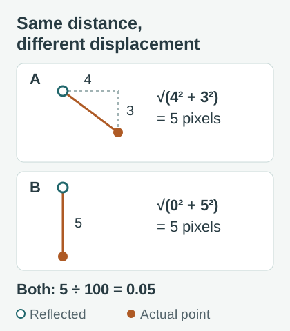
Even the geometry has a limit: a point displaced four pixels sideways and three vertically has the same five-pixel residual as a point displaced five pixels vertically. With 100-pixel reference spacing, both give 5 ÷ 100 = 0.05, a normalized mismatch for one landmark pair. The full checker averages multiple pairs. Equal distance does not mean the same movement, tissue or cause.
If you are exploring a long-standing appearance without new symptoms, use one clear frontal photo with even light and a relaxed expression. Keep an original. There is no need to repeat scans until a preferred result appears. Read the measurement method and why scores vary.
Bring a short history, not a score target
For a non-emergency visit, write these observations in a note on your phone or on paper. Do not delay urgent care to complete the list.
- Timing: when you first noticed it, whether onset was sudden or gradual, and whether it is changing.
- Location: describe the feature and name your own left or right side; mirrored photos can reverse the display.
- Function: any pain, altered sensation, swelling, chewing difficulty, bite change, speech change or trouble closing an eye.
- Context: recent dental work, injury, procedures, illnesses or medicines to discuss with the clinician.
- Images: a small number of dated originals, if already available. Avoid filters or mirrored composites.
Ask, “What findings support the explanation? Is an examination or test needed? What change should prompt earlier follow-up?” A dentist can assess tooth and bite concerns; a medical clinician can assess other new symptoms and arrange referral when needed.
Common questions about an uneven face
Why is one side of my face bigger?
“Bigger” might describe the jaw outline, cheek fullness, a muscle, swelling or a photographic effect. Those are different observations. Note the location, timing and any symptoms. New, persistent or increasing fullness should be assessed rather than assigned a cause from a mirror comparison.
How can I tell whether it is bone, muscle or fat?
A normal selfie cannot reliably separate these. A clinician may examine shape, movement, teeth and bite and decide whether imaging is needed. The jaw symmetry photo tool compares visible landmarks; it cannot identify the tissue responsible or determine whether jaw growth is active.
Does jaw clicking explain my facial asymmetry?
Clicking alone cannot establish a cause. Painless jaw-joint clicking is common and does not itself need treatment, according to NIDCR. Tell a dentist about pain, locking or movement difficulties rather than diagnosing yourself from a photograph.
Can facial asymmetry be natural and still deserve assessment?
Yes. Small long-standing differences are common, yet “natural” is not a clinical diagnosis. Ask for assessment when a difference is new, progressing, painful or affects function, or when you are unsure about its history. A high photo score cannot rule out a health problem.
Will facial exercises or massage fix the cause?
An appearance alone does not identify what needs treatment. Evidence about an association with a habit does not validate an exercise, massage or chewing program for an individual. Specific rehabilitation may be appropriate for a diagnosed condition under professional guidance. See the guide to options and their limits.
Sources and editorial review
- Penn Medicine: Facial asymmetry - clinical overview of contributors and assessment.
- Johns Hopkins Medicine: Hemifacial microsomia - one defined developmental condition.
- Linden et al. (2018): The Relationship between Age and Facial Asymmetry - 3D measurements across 191 volunteers.
- Harnádková et al. (2024): Facial directional asymmetry and laterality - observational 3D study of 163 adults.
- Heikkinen et al. (2024): Chewing Side Preference, Facial Asymmetry and Related Factors - 748 adults; no significant relationship between chewing preference and facial asymmetry.
- Liu et al. (2014): Factors contributing to facial asymmetry in identical twins - habit associations in 147 twin pairs, not a sleep intervention.
- NIDCR: Temporomandibular disorders - symptoms, diagnosis and treatment limits.
- NHS: Stroke symptoms · Bell’s palsy · Dental abscess - urgent symptoms and assessment.
Published by Face Symmetry Checker. Content and sources checked . This is general educational information, with no independent clinical review claimed. Diagrams are original teaching illustrations, not patient images. Read our editorial policy.