
How Our Face Symmetry Test Works
Follow one photograph from estimated landmarks to its displayed result. This method explains the selected pairs, eye-line reference, normalization, regional weights and score calculation, alongside the checks performed and the validation still needed.
From image pixels to estimated points
The automatic checker accepts a supported photo or captures camera frames with your permission. A large uploaded image is resized proportionally so its longest side is at most 1,600 pixels. All distances then refer to that working image. A distance in pixels cannot be converted into millimeters without a separate, valid calibration.
The model files load from this website when analysis starts. Selected-photo inference runs in the browser, normally in a worker; a main-thread fallback is available. This avoids an image-upload backend for the analysis. It still requires ordinary website and model-file requests. Read the privacy page for that distinction, and remember that a downloaded image becomes a separate file you control.
We use MediaPipe Face Landmarker float16 v1 with @mediapipe/tasks-vision 1.0.1. Google documents a 478-landmark output, expression coefficients and transformation matrices. The checker uses estimated x/y coordinates for the measurement, expression coefficients for limited warnings and the matrix for pose checks. The model’s estimated depth is not used to claim a three-dimensional reconstruction or perspective correction.
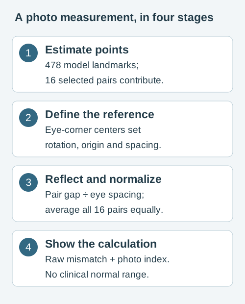
The eye line defines alignment and scale
The eye centers here are the midpoints of the inner and outer eye corners: landmarks 33 and 133 on one side, 263 and 362 on the other. They are not detected pupil or iris centers. Their midpoint is the origin; the line joining them supplies the rotation used to level the photo coordinates. Eye spacing, d, is their Euclidean distance before that rotation.
Normalized x is multiplied by the working image width and normalized y by its height before any distances are calculated. Using x/y fractions directly on a rectangular image would distort the geometry. After translation and rotation, the comparison line is the vertical line through the eye-center midpoint. It is a reproducible reference convention, not a clinically established facial midline.
For each image point P:
Q = rotate(P − eye-center midpoint, −eye-line angle)
reflection of Q = (−Qx, Qy)
Leveling absorbs the relative height difference between the two eye centers into the reference. This automatic index therefore cannot independently report that same eye-height difference. The manual eye symmetry tool leaves the photograph’s horizontal axis in place and answers a different question.
For fixed landmarks, translation, in-plane rotation and uniform scaling do not change the normalized pair distances. A horizontal image reversal preserves the geometry when the corresponding point labels are swapped. Changing camera viewpoint is different: yaw, pitch, perspective, expression and new model estimates can change the points. Alignment cannot undo those effects.
The 16 selected landmark pairs
The detector can estimate many points, but only the following pairs contribute to this index. IDs are zero-based model indices. Feature names describe their approximate location on the visible photo; a contour point is not a measurement of the underlying bone. Image-left and image-right labels describe the view, rather than assuming how a camera saved or mirrored the image.
| Region | Visible feature | Landmark IDs |
|---|---|---|
| Eyes | Outer eye corners | 33 ↔ 263 |
| Eyes | Inner eye corners | 133 ↔ 362 |
| Eyes | Upper eyelid points | 159 ↔ 386 |
| Eyes | Lower eyelid points | 145 ↔ 374 |
| Eyebrows | Outer brow contour | 70 ↔ 300 |
| Eyebrows | Central brow contour | 105 ↔ 334 |
| Eyebrows | Inner brow contour | 107 ↔ 336 |
| Nose | Nose-base side points | 98 ↔ 327 |
| Nose | Lateral nose-area points | 129 ↔ 358 |
| Mouth | Mouth corners | 61 ↔ 291 |
| Mouth | Upper lip contour | 40 ↔ 270 |
| Mouth | Lower lip contour | 91 ↔ 321 |
| Cheeks | Lateral face contour | 234 ↔ 454 |
| Jaw | Lower lateral face contour | 172 ↔ 397 |
| Jaw | Lower face contour | 136 ↔ 365 |
| Jaw | Chin-adjacent contour | 149 ↔ 378 |
Equal pair weights produce unequal region weights
Each pair contributes one-sixteenth of the total mean. The eyes have four pairs (25% of the mean); eyebrows, mouth and jaw each have three (18.75% each); the nose has two (12.5%); cheeks have one (6.25%). The overall result is not the unweighted average of the six region summaries. These weights follow the selected point set; they are not claims about clinical importance or attractiveness.
A region’s own mismatch is the mean of its pairs. You can inspect the individual rows in the checker instead of accepting the headline number alone. A sparse selected-point map does not measure every patch of skin, muscle movement, texture or facial volume.
Reflect, measure the gap, then normalize
For each aligned pair, reflect the first point across the vertical comparison line and measure the straight-line distance to its counterpart. The distance includes horizontal and vertical differences together; their Euclidean combination prevents one component from canceling the other.
pair gap = √[(−Qax − Qbx)² + (Qay − Qby)²]
pair mismatch = pair gap / d
A = sum of the 16 pair mismatches / 16
mismatch (% of eye spacing) = 100 × A
photo symmetry index = max(0, 100 − 100 × A)
All mismatches are nonnegative, so the index cannot exceed 100. The zero floor prevents a negative display value when the mean exceeds the reference spacing. This transform is a product convention, rather than an independently calibrated “symmetry percentage.”
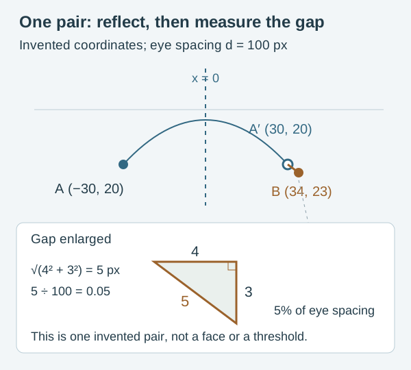
A worked example you can reproduce
Let eye spacing be 100 pixels. Point A at (−30, 20) reflects to (30, 20). Point B is at (34, 23). The gap is √(4² + 3²) = 5 px, so this pair’s mismatch is 5 / 100 = 0.05, or 5% of eye spacing.
If all 16 invented pairs had that same gap, the mean would be 0.05 and the index 95. One pair by itself cannot determine the overall score. An index of 95 does not mean that 95% of a face is symmetrical, nor does it describe a typical person.
On screen, the headline index is rounded to a whole number; detailed pair values have their own display rounding. JSON preserves numerical values before display rounding. Recomputing a mean from rounded table values can therefore differ slightly from the downloaded result.
How the manual photo tools measure their points
The feature tools use points you select in the working photo. They do not run the automatic detector or use its 16-pair score. A pixel distance is measured in the resized working image, not millimeters of anatomy. Percentages below name their own reference distance; they are not percentages of a face that is symmetrical.
| Tool and references | Calculation and units |
|---|---|
| Eye symmetry: four eye corners | Each eye's reference center is the midpoint of its selected inner and outer corners. Height difference is the absolute image-y difference between those midpoints divided by their Euclidean spacing, times 100. Eye width is corner-to-corner Euclidean distance; the ratio is image-left width / image-right width. |
| Canthal tilt: the same four corners | atan2(inner y − outer y, |outer x − inner x|), in degrees for each eye. Positive means outer corner higher. Difference is the absolute difference between the two angles. Both use image horizontal, with no automatic roll alignment. |
| Smile: two corners and a selected philtrum center | Corner-height difference, reflected-corner distance and horizontal half-width difference are each divided by the corner-to-corner Euclidean distance, times 100. Reflection uses a vertical line through the selected philtrum center. Only its x-coordinate defines this line; moving that center up or down without changing x does not alter these three values. |
| Jaw and nose: bridge/base axis, two outline points and chin or tip | Reflect the image-left outline point across the selected bridge-to-base axis. Divide its distance to the other outline point by their Euclidean width, times 100. Chin/tip offset is the absolute perpendicular distance to that axis divided by the same width, times 100. The tool also separates reflected mismatch into side-distance difference (unequal perpendicular distances to the axis) and along-axis level difference. Both use that width and their hypotenuse equals the reflected mismatch. These components describe the selected points, not additional anatomical measurements. |
| Thirds and fifths: four or six ordered boundaries | Project the boundaries onto the first-to-final reference axis. Divide each consecutive projected interval by the total axis length, times 100. The segment shares sum to 100%; the endpoints define that total. |
| Photo proportions: two endpoint pairs | Calculate the Euclidean length of A and B in working-image pixels, then A / B. The eye-spacing preset uses selected iris centers; the automated checker and manual eye tool instead use eye-corner midpoints. |
| Golden-ratio calculator: two positive entered lengths | Divide same-unit A by B and compare the dimensionless ratio with φ ≈ 1.618. It does not choose facial endpoints or infer appearance from that reference. |
Which eye center and which reference axis?
An iris center, a pupil center and the midpoint of two eye corners are different definitions. The manual eye tool's center is a corner midpoint; it does not locate the pupil or measure eyelid opening. The automatic checker levels the line between detected corner midpoints, absorbing their relative image-height difference. The manual eye tool leaves that image-relative difference visible. These methods can give different outputs for the same tilted photo.
The jaw and nose axis is a reference you choose from visible nose points. It is not a calibrated anatomical midline. Nose-wing edges describe the outer contour, not the openings of the nostrils or the internal septum. Moving an axis point changes both the reflected mismatch and perpendicular offset.
For thirds, the glabella is the selected central area between the eyebrows, rather than an average eyebrow-height line. The visible nose base and bottom of chin are photo references. Hairline placement and the selected face outlines can change the denominator. These choices should remain consistent when comparing images.
Manual point placement, perspective, head roll and expression all affect the results. Neither the manual values nor the detector's index has a validated clinical normal range or attractiveness threshold.
The two-photo comparison is different again: it runs the same automatic method separately for A and B and reports B’s mean mismatch minus A’s in percentage points of eye spacing. The exported pair components satisfy horizontal² + vertical² = reflected mismatch². A positive difference means greater mismatch in B under this method; it does not identify a treatment effect.
The mirror generator, horizontal flip and portrait preference tool perform image or choice workflows. They do not add extra measurements to the 16-pair index.
What the photo checks can and cannot establish
The automatic workflow requires one detected face, finite coordinates inside the image, and at least 60 working-image pixels between eye centers. Estimated pose checks reject an absolute yaw over 12 degrees or pitch over 22 degrees; an unavailable or invalid pose matrix also prevents acceptance. These are implementation heuristics, not clinically validated acceptance ranges.
Expression coefficients generate a warning when jaw-open exceeds 0.30 or either smile coefficient exceeds 0.55. Missing expression data is disclosed rather than assumed neutral. Cheek samples provide brightness warnings: their relative difference exceeds 0.35, or their mean is below 40 on the sampled 0–255 luminance scale. These numbers do not establish that a face is evenly illuminated or correctly detected.
A photo can pass and still contain inaccurate points. Glasses, hair, blur, shadows, cosmetics, facial hair, filters and model limitations can matter. Examine whether the overlay fits the visible features; use a neutral expression, visible contours and similar camera conditions when comparing photos. Camera perspective and score variation explain why a new photo is a new input.
How five-frame camera scans are summarized
A camera scan collects five accepted frames, measuring each independently. It may attempt up to 15 frames to obtain them. The headline index is the median of the five frame indices. The headline mismatch is the median of their mismatches; because there are five values and the transform is monotonic, these summaries correspond under the published formula.
The displayed photo, pair rows and regional details come from the accepted frame closest to the median mismatch. Under the present five-frame method, that is a median-mismatch frame. The capture chart and JSON list the observed frame mismatches. The minimum and maximum describe that short capture only. They are not a confidence interval, an uncertainty estimate for anatomy or a validated day-to-day repeatability band.
Nearby frames share camera, pose, lighting and detector behavior. A narrow range can coexist with a consistently misplaced landmark. Avoid repeating scans until you obtain a preferred number. If your goal is to compare two photos, keep the method and capture conditions consistent and retain the raw mismatch alongside the index.
Validation status and accuracy limits
What we check in software
Constructed-coordinate checks cover the equation, weighting, scale and rotation behavior, invalid inputs and selected manual geometry. Browser checks exercise supported uploads, sample inference, overlays, downloads and clearing. These checks can catch implementation errors and misleading descriptions.
What has not been established
We have not conducted a consented human accuracy study against a suitable reference, a representative population study or clinical validation of this index. Physical-device and demographic coverage also need further evaluation. No accuracy percentage, normal score band or meaningful-change cutoff has been established.
Adding more points, unequal weights or model-estimated depth would change the measured quantity. Such changes need a stated purpose, evidence and a versioned method; they cannot simply be called “more accurate.” The current math already accounts for both axes and image dimensions. Its main unresolved question is the relationship between these selected photo estimates and an appropriate external reference.
A high index cannot determine health, attractiveness, identity, treatment need or self-worth. New facial weakness is a separate health question: follow the urgent guidance and do not delay care to measure a photo.
Questions about the measurement
Why use eye spacing instead of face width?
Eye-corner centers provide one explicit scale and alignment reference for this point set. Face-outline width would be another convention and would change the denominator. Neither choice creates a calibrated anatomical scale. The selected reference must remain consistent when comparing measurements.
Can I reproduce my result from the JSON?
For a single photo, average the 16 full-precision mismatchPercentOfEyeSpacing values in landmarkPairs. That mean should match mismatchPercentOfEyeSpacing; subtract it from 100 and apply a zero floor to reproduce score. Small floating-point differences can occur. For a camera scan, also read cameraFrameMismatchPercent and the median-frame explanation above.
Why does moving the mirror line leave the index unchanged?
The mirror-line control changes the visual composite. Numerical alignment stays anchored to the detected eye-center reference. This keeps an image-editing choice from silently changing the score. The exported visualMirror field records the visual offset separately.
Is the model’s confidence an accuracy percentage?
No. Detection and presence settings govern whether the model returns a face; they do not quantify the anatomical error of the landmarks or validate our index. Passing a pose or brightness rule is also different from establishing measurement accuracy.
Why are other apps’ scores different?
They can choose different detectors, points, axes, scales, weights and display transforms. Even two numbers out of 100 can describe different quantities. Our app comparison guide starts with the output you need rather than a score ranking. Compare the tool types.
Sources, implementation and review notes
The point set and formulas above document this website’s implementation. The external references support detector definitions and photographic context; none independently validates this product. Content and code correspondence checked . Diagrams are original invented teaching examples, not patient images.
- Google: Face Landmarker for Web - outputs, configuration and worker considerations.
- Google: NormalizedLandmark interface - x/y reference dimensions and model-estimated depth.
- MediaPipe canonical face model - technical mesh reference, not a beauty template.
- Ward et al. (2018): Nasal Distortion in Short-Distance Photographs - a perspective model, not a correction formula for our score.
Published by Face Symmetry Checker. Read the editorial policy or report a method or explanation issue. We do not claim independent clinician review.
Reproduce the head-turn sensitivity experiment
A symmetric reference model can produce a lower 2D index when turned. Our reproducible pose-sensitivity experiment shows the calculation, assumptions and data. It tests the formula’s response to a known projection; it does not validate real-face accuracy.